
Figure 3.
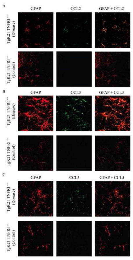
Confocal microscope image of the brains of TgK21 mice immunolabeled with antibodies against the astrocyte marker GFAP and CCL2 (A), CCL3 (B) and CCL5 (C). Note marked increase in GFAP immunoreactivity in TNFαR+/+ mice that were commonly clinically compromised. Coronal brain sections were at the middle portion of the parietal lobes. Photomicrographs are representative of two separate experiments with each experiment having 3 mice per group.