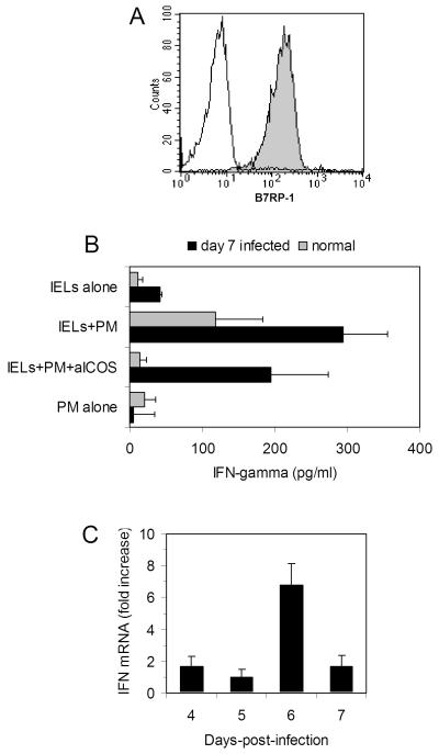
FIG. 6.
(A) The B7RP-1 molecule, the ICOS ligand, is expressed (shaded area) at high density on PM cells; control staining (unshaded area). (B) IFN-γ secretion by IELs from normal mice (grey bars) or mice 7 days post-infection (black bars) following 24 culture either alone, with the PM cells, or with PM cells in the presence of anti-ICOS mAb. Although culture of IELs from normal and virus-infected mice resulted in an increase in IFN-γ secretion, IFN-γ was consistently higher from infected mice. Treatment of IELs with anti-ICOS mAb inhibited IFN-γ responses of both normal and virus-infected mice, indicating that stimulation of IELs by PM cells occurs through an ICOS-mediated signal. Data are mean values ± SEM of 5 normal mice and 4 virus-infected mice. (C) Real-time PCR analysis of IFN-γ gene expression in CD4+ cell-sorted IELs from days 4, 5, 6, and 7 post-reovirus infection. Data are mean values of two samples expressed as fold increase over non-infected mice using the 2-ΔΔCt method [27] as described in the Materials and methods.

