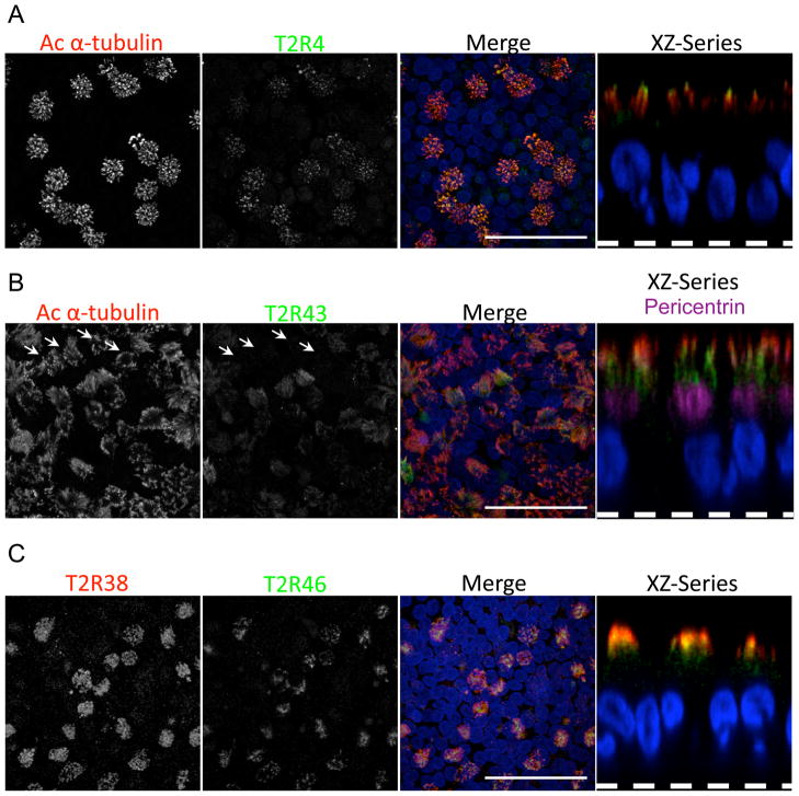
Figure 2.
T2Rs localize along the motile cilia of airway epithelia. Confocal immunofluorescence microscopy of cultured human airway epithelia with anti-T2R antibodies showed that (A) T2R4 and (B) T2R43 (both green) localize to motile cilia, which were identified by antibody to acetylated α-tubulin (red). Nuclei stained with DAPI are blue. Arrows in panel B indicate location of ciliated cells that were not labeled by anti-T2R43 antibody. (C) T2R38 (red) and T2R46 (green) localize to motile cilia. Data are stacks of confocal z-series images in X-Y plane and a single X-Z plane image (right, dashed line indicates the filter). Panel B (right) also shows anti-pericentrin antibody (purple), which labels the basal body below cilia. Scale bars indicate 20 μM. See also Fig. S1–S7.

