Figure 3.
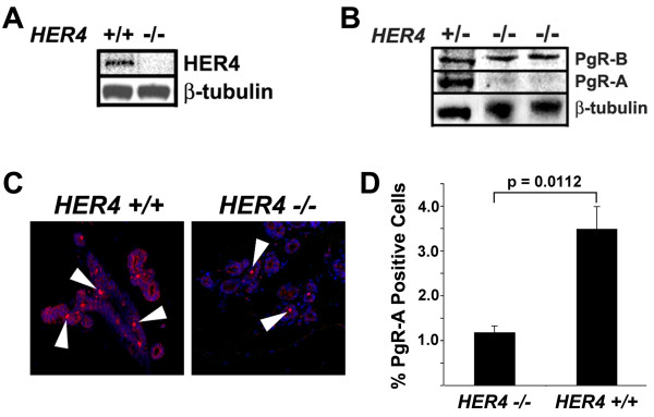
HER4 regulates PgR expression in the mouse mammary gland. (A) Transgenic mice expressing HER4 under the control of the cardiac specific myosin promoter were crossed with HER4+/- mice (both kindly provided by Jon Golding, Open University) to generate HER4-/-;HER4heart (HER4-/-) mice [17]. Mammary glands were collected and snap frozen in liquid nitrogen or spread on a glass slide, fixed overnight in formalin, processed, and paraffin embedded. Lysates were prepared from P14.5 mammary glands and analyzed by western blot using an antibody that recognizes (A) HER4 (Cell Signaling 111B2) or (B) both PgR-A and PgR-B isoforms (DAKO A0098). (C) Immunofluorescent localization of PgR-A in mammary glands at P14.5. After antigen retrieval by pressure cooker treatment in citrate buffer, sections were incubated in peroxidase block followed by DAKO block before overnight incubation with the PgR-A specific anti-PgR sc-538 (Santa Cruz) [18] antibody. Secondary antibody was Alexa 555-conjugated (Invitrogen) and nuclei were counterstained with DAPI. Arrowheads indicate nuclear staining of PgR-A. (D) The number of PgR-A-positive alveolar cells were quantitated from images captured using a TCS SP2 Leica confocal microscope with an Acousto-Optical Beam Splitter. Mammary glands from three mice per genotype were analyzed and a minimum of 1000 alveolar cells per mouse were counted. Results are expressed as mean +/-SEM. Statistical significance was determined using the Student's t test. A significant (p = 0.0112) 2.95-fold reduction in PgR-A positive alveolar cells was observed in mammary glands lacking HER4 expression when compared to wild-type control mammary glands.

