Figure 4.
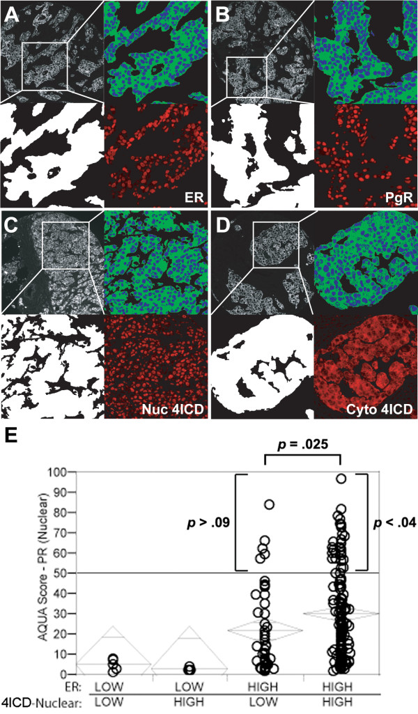
Nuclear 4ICD coactivates PgR expression in human breast carcinomas. A cohort of 196 samples of PgR(+) invasive ductal carcinomas from the Yale University Department of Pathology diagnosed from 1961 to 1983 were stained in a tissue microarray format using a modified indirect immunofluoresence method and antibodies described previously [20] with the addition of anti-PgR; PgR636 (Dako) and anti-HER-4 sc-283 (Santa Cruz Biotechnology, Santa Cruz, CA) antibodies. Subcellular immunofluoresence was analyzed by a pathologist using the AQUA™ method as published previously [20,21]. (A-D) Immunofluorescent illustration of AQUA®. In each panel, representative pseudo-colored images are shown of cytokeratin (upper left), tumor mask (lower left), nuclear (blue) and non-nuclear or membrane compartments (green) (upper right), and target expression (red) after RESA application (lower right) as described elsewhere [20,21]. (A) ER nuclear expression, (B) PgR nuclear expression, (C) nuclear expression of 4ICD, (D) predominantly cytoplasmic expression of 4ICD. (E) AQUA scores for ERα and nuclear 4ICD were dichotomized to examine the association of these markers with continuous PgR AQUA scores. The cutpoint for ERα was chosen as 3.75 (bottom 25% vs. top 75%) [1]. The cutpoint for nuclear 4ICD was near the cohort median at 10. Chi-square analysis revealed a significant association between PgR and ERα expression (p < .0001). The PgR AQUA score was significantly greater in tumor samples with ERα and nuclear 4ICD coexpression (p = .025). The highest levels of PgR expression (AQUA > 50) were significantly associated with ERα and nuclear 4ICD coexpression (p < .04).

