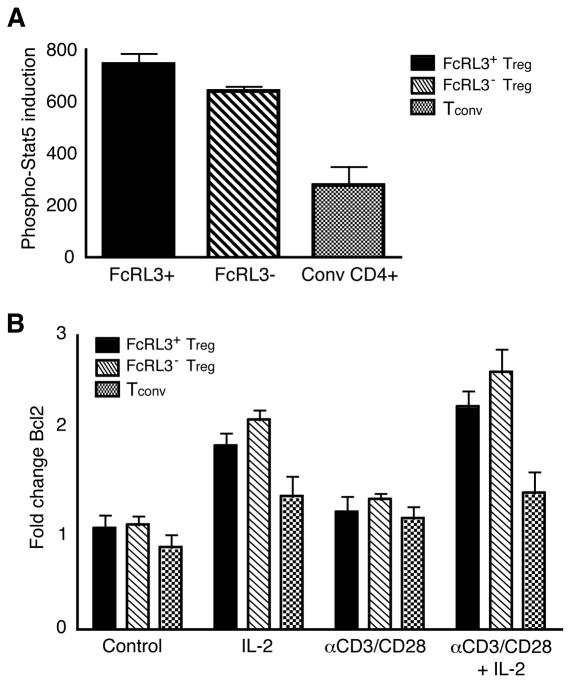
Figure 6. FcRL3+ and FcRL3− Treg demonstrate intact proximal and distal signaling responses to IL-2.
A. FACS-sorted FcRL3+ Treg, FcRL3− Treg, and Tconv were incubated with either medium alone or IL-2 for 30 minutes, fixed, and then stained for Stat-5 using an antibody specific for the phosphorylated Y694 Stat-5 epitope. The specific induction of phospho-Stat-5 is shown (MFI after IL-2 stimulation minus MFI after R-10 medium alone); data represent mean +/− SEM of 3 separate experiments comprising 3 different donors. B. FACS-sorted FcRL3+ Treg, FcRL3− Treg, and Tconv were cultured in the presence of medium alone, IL-2, αCD3/CD28 antibodies, or a combination of αCD3/CD28 antibodies and IL-2. After 5 days, cells were fixed, permeabilized, and stained for Bcl2. The fold change in Bcl2 expression from day 0 to analysis at day 5 is shown; data represent mean +/− SEM of 3 separate experiments comprising 4 different donors.

