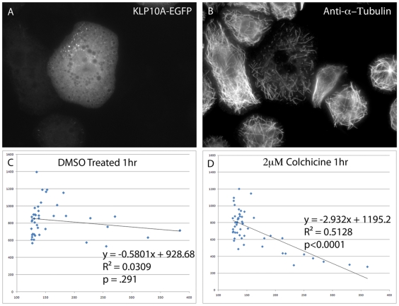
Figure 4. Overexpression of KLP10A-GFP in Drosophila S2 cells confers increased susceptibility to colchicine treatment.
S2 cells were transfected, induced to express KLP10A-GFP, treated with 1 µM colchicine for 1 hour, then processed for immunofluorescence to visualize microtubules. A representative transfected cell is shown in the tubulin channel (A) and GFP channel (B). Note extensive loss of microtubules in (A). In order to test for a correlation between expression levels of KLP10A-GFP and the extent of microtubule depolymerization, individual transfected cells were imaged in both channels and microtubule polymer levels and KLP10A-GFP expression levels were determined by measuring average fluorescent pixel intensities per cell. Bar = 5 µm. The results are plotted for control DMSO-treated cells (C) and colchicine-treated cells (D). The amount of KLP10A-GFP in DMSO treated cells (C) did not show a statistically significant effect on the amount of MT polymer present (p = 0.291). However, when transfected cells were treated with colchicine (D) a strong negative correlation is observed between the amount of KLP10A-GFP present and the amount of MT polymer present (p<0.0001).

