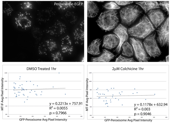
Figure 5. GFP expression alone does not protect microtubules from colchicine.
As a negative control, S2 cells were transfected with a plasmid expressing GFP targeted to peroxisomes, either treated with DMSO or colchicine, and processed as described in Figure 3 to visualize microtubules (A) and peroxisomes (B). Bar = 5 µm. The plots show average microtubule pixel intensity per cell plotted against GFP peroxisome average pixel intensity per cell. The amount of Peroxisome-GFP did not show a statistically significant effect on the amount of MT polymer present in DMSO-treated cells (p = 0.7966, C) or colchicine-treated cells (p = 0.9046, D).

