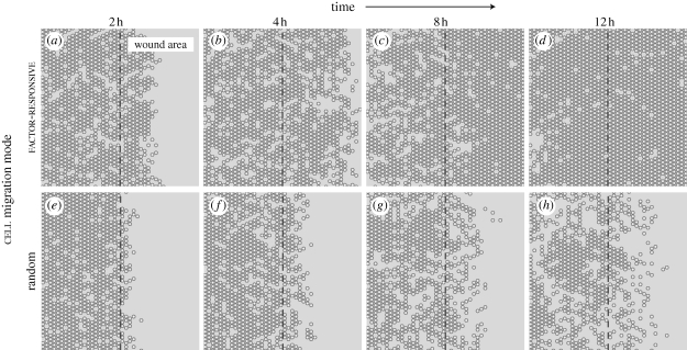
Figure 4.
Simulated AT II cell monolayer wound healing. In scratch wound healing assays, a confluent cell monolayer is scratched with a probe to inflict a denuding wound. Following injury, surviving cells move into the denuded area and eventually restore monolayer confluence. We simulated that protocol by creating a confluent cell monolayer, and then removing contiguous columns of cells across the middle. We explored two different cell migration modes: random and factor-responsive. (a–d) Shown are simulated wound healing images of factor-responsive cells, which migrated away from a wound-induced, diffusive factor. Objects with white centres are cells. The dashed line indicates wound margin; to its right is the denuded area. As simulation progressed, an increasing number of cells migrated into the wound area and closed most of the wound within 12 h post-wounding. One simulation cycle mapped to approximately 35 min in vitro. (e–h) Cells migrating randomly exhibited poor wound healing characteristics, and failed to close the wound within 12 h.

