Figure 5.
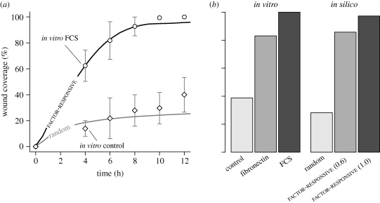
Simulated and in vitro wound closure of AT II cell monolayer. See Kheradmand et al. (1994) and Garat et al. (1996) for details on in vitro assays and wound healing data. Briefly, AT II cell monolayers grown in the presence of different factors such as FCS and fibronectin were scraped once with a 23-gauge probe, and images captured at regular intervals over a 24 h period. Wound width was 50 cell-widths, which maps to 0.5 mm in vitro. We used two migration modes: random and factor-responsive. In factor-responsive mode, cells migrated away from a wound-induced factor. (a) Shown are wound closure of two in vitro conditions, and simulated wound healing using the two migration modes. Factor-responsive cells closed wounds at rates comparable to the in vitro condition with FCS, and regained monolayer confluence by 12 h. Randomly migrating cells failed to close the wound, and exhibited kinetics somewhat similar to the in vitro control condition. One simulation cycle mapped to approximately 35 min in vitro. Bars: ± s.d. (b) Shown are in vitro and simulated wound closures after 24 h. Compared with FCS, fibronectin was a less potent wound-healing agent. Cells migrating at 1.0 grid unit per cycle closed 97% of the wound. Reducing cell speed to 0.6 grid units per cycle led to 86% partial closure, which roughly matched the in vitro condition with fibronectin. The simulation data represent mean values of 100 Monte Carlo runs.

