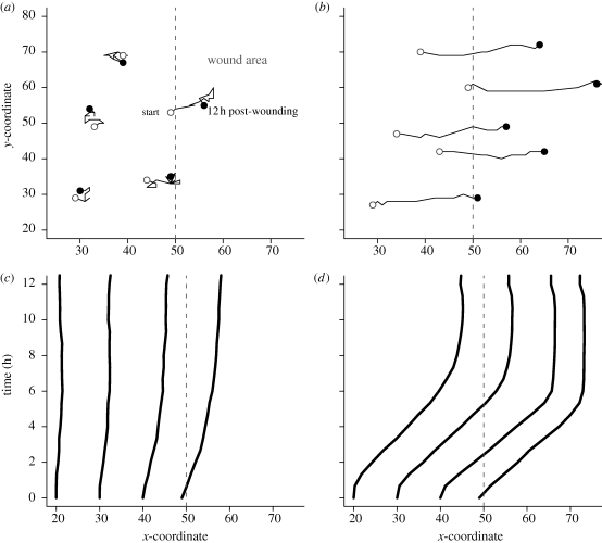
Figure 6.
Cell movement tracked during simulated wound healing. Simulations are the same as in figure 5. (a,b) Shown are typical movements for 12 h post-wounding by cells migrating (a) randomly or (b) directionally. Randomly migrating cells closer to the wound margin (dashed line) initially exhibited semi-persistent directionality towards the wound area, but soon lost the direction. Factor-responsive cells had persistent trajectories towards the wound area. (c,d) Shown are patterns of cell displacement from their initial positions. (c) Displacement of randomly migrating cells at specific distances away from the wound margin was small. Cells at the wound margin showed modest displacement over the 12 h period. (d) Factor-responsive cells had large displacements, which were consistent regardless of the initial distance away from the wound margin. Each curve represents mean displacement of cells initially at the same distance away from the wound.

