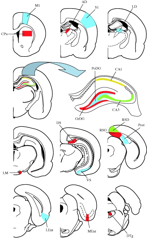
Figure 2.
Diagrams of coronal sections showing the location of the 18 brain regions investigated. Schemes adapted from Paxinos & Watson (2005). AD, anterodorsal thalamic nucleus; CA1, field CA1 of the hippocampus; CA3, field CA3 of the hippocampus; CPu, caudate putamen (striatum); DS, dorsal subiculum; DTg, dorsal tegmental nucleus; GrDG, granular layer of the dentate gyrus; LD, laterodorsal thalamic nucleus; LEnt, lateral entorhinal cortex; LM, lateral mammillary nucleus; M1, primary motor cortex; MEnt, medial entorhinal cortex; PoDG, polymorph layer of the dentate gyrus; Post, postsubiculum; RSD, retrosplenial dysgranular cortex; RSG, retrosplenial granular cortex; S1, primary somatosensory cortex; VS, ventral subiculum.

