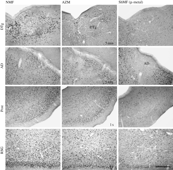
Figure 4.
Brightfield photomicrographs showing c-Fos immunoreactivity in brain regions that contain the head direction cells. Animals were exposed to the different magnetic conditions while nesting in an unfamiliar circular arena. AD, anterodorsal thalamic nucleus; AZM, experimental magnetic field that was shifted in azimuth by 120° every 5 min or every second (as indicated in the photomicrographs); DTg, dorsal tegmental nucleus; NMF, natural geomagnetic field; Post, postsubiculum; RSG, retrosplenial granular cortex; ShMF (µ-metal), shielded magnetic field. Scale bar, 300 µm.

