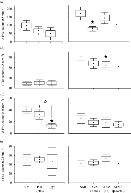
Figure 8.
Number of c-Fos-IR cells in the entorhinal cortex and the subiculum. Note that only a single independent datum point was available for the MEnt, LEnt and VS under shielded magnetic field conditions (ShMF) (cf. §2.4.). See caption of figure 3 for explanation. Closed asterisks indicate comparison with a control group of animals exposed to the local geomagnetic field, open asterisks comparison with another experimental group. (a) Medial entorhinal cortex (MEnt); (b) lateral entorhinal cortex (LEnt); (c) dorsal subiculum (DS); (d) ventral subiculum (VS).

