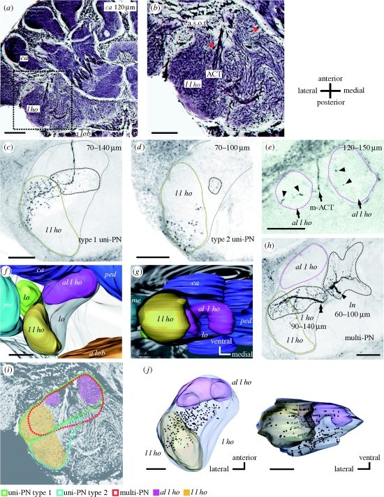
Figure 1.
Structure of the l ho of the ant brain. (a) A frontal section of a Bodian-stained ant brain at a depth of approximately 120 μm from the frontal (ventral) surface. a lob, antennal lobe; l ho, lateral horn; ca, calyces of the mushroom body. (b) A magnified image of the lateral protocerebrum (inset in (a)), including the l ho. Two round structures at the anterior part of the l ho (red arrowheads), which we call antero-lateral l ho (al l ho), are delineated by the antenno-cerebral tract (ACT) and the anterior superior optic tract (a.s.o.t). In addition, an ellipsoidal structure at the lateral edge of the l ho, lateral l ho (l l ho), is also visible. (c) Terminal arborizations of a class of uni-PN (type 1 uni-PN), the axon of which passes through the medial antenno-cerebral tract (m-ACT), seen as a confocal image at a depth of 70–140 µm from the frontal surface. Their terminal branches are located in the anterior part of the lateral l ho (region denoted by yellow broken line) and nearby l ho region (region denoted by black broken line). Grey broken line in (c), as well as in (d) and (h), depicts outline of the l ho at this depth. (d) Terminal arborizations of another class of uni-PN (type 2 uni-PN). This type terminates in the posterior part of the lateral l ho (yellow broken line) and nearby l ho region (black broken line). (e) A type 1 uni-PN with terminal arborizations (arrowheads) in the antero-lateral l ho (magenta broken lines). (f,g) A three-dimensional reconstruction of the lateral l ho, and the antero-lateral l ho, and nearby protocerebral neuropils, viewed (f) ventrally and (g) posteriorly. The antero-lateral l ho is positioned postero-laterally to the mb, and the lateral l ho is positioned postero-medially to the medulla (me) and lobula (lo). ped, pedunculus. (h) The axon of a multiglomerular PN (multi-PN) bifurcates (arrowhead and double arrowhead), each forming dendritic branches in the l ho and in the area called the lateral network (ln) (Kirschner et al. 2006; Zube et al. 2008). (i) A schematic diagram of termination areas of type 1 uni-PNs (green), type 2 uni-PNs (blue) and multi-PNs (red) in the l ho. (j) Three-dimensional reconstruction of the distribution of terminal boutons of a multi-PN in the l ho, viewed ventrally and posteriorly. Scale bars: (a) 100 μm; (b–h, j) 50 μm.

