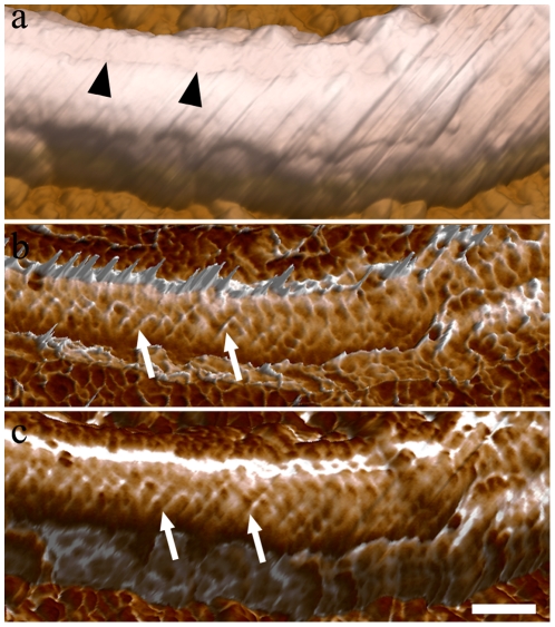
Figure 5. AFM intermittent contact mode image of bending state flagellum of detergent-extracted epimastigote forms.
(a) Topographic 3D view of the PFR structure. The furrow can be observed (arrowheads). (b) Phase image of the flagellum in a bending state shows the distribution of the filaments of the PFR (arrows) along the flagellum. (c) 3D view of combined height and phase images. Bar – 200 nm.

