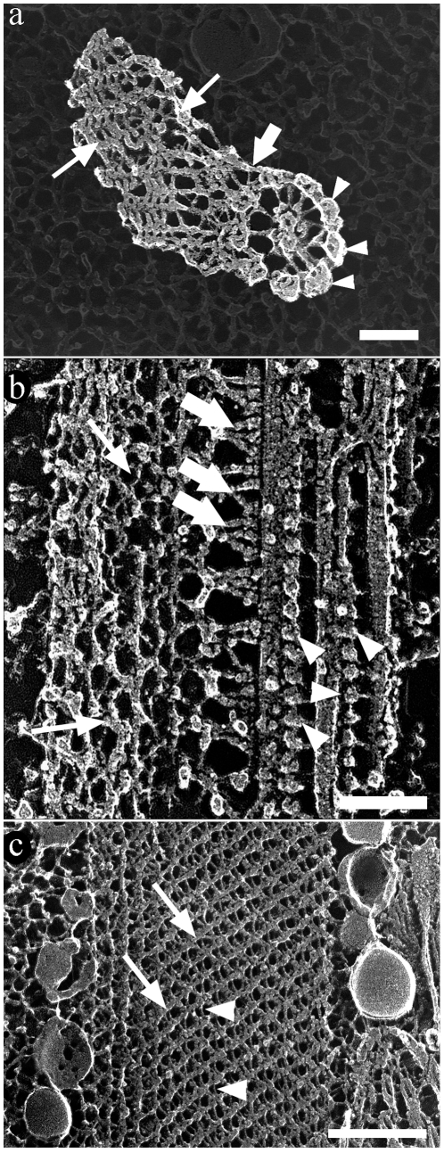
Figure 6. Deep-etching replica images of the flagellum of T. cruzi epimastigote form.
The main components of the flagellum are clearly shown in transversal (a) and longitudinal (b–c) fractures. (a) The peripheral (arrowheads) as well as the central pair of microtubules of the axoneme are seen. The PFR is seen in the left side of the figure (arrows). (b) Transversal fracture of the flagellum shows microtubules of the axoneme, dynein units (arrowheads) and the PFR (arrows). The flagellum is in a straight state with the PFR in a relaxed state. (a–b) The axoneme and the PFR are linked by fibers indicated by thick arrows. (c) Deep-etching replica image of the PFR with intercrossed thicker (arrows) and thinner filaments (arrowheads). These images were digitally processed in order to emphasize only the components of the flagellum against a shallow background. Bar – 100 nm.

