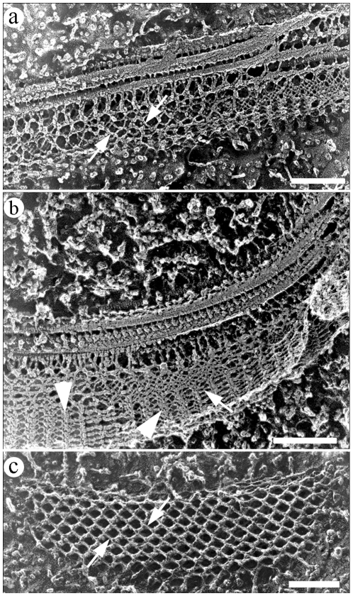
Figure 7. Deep-etching replica of flagellar regions in different states: straight (a) and bent (b).
Angles between PFR filaments (arrows) vary according to the bending state of the flagella. (c) Deep-etching image showing a longitudinal fracture of the intermediate domain of the PFR in a flagellum presumably in a straight state. Band-like structures on (b) are indicated by arrowheads. Bars – 250 nm.

