
Fig. 4.
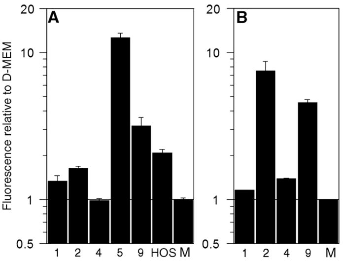
Proteolytic activity of (A) MMP-1 and (B) MMP-13 in concentrated media conditioned by HOS or GCT stromal cells for 24 h, as determined by protease-specific standardized activity assays and relative to serum-free D-MEM (“M”). Quantification was achieved through measurement of fluorescence following incubation with a quenched fluorophore that fluoresced upon proteolytic cleavage by MMP-1 or MMP-13. GCT cell lines tested are indicated numerically and refer to GCT-1, -2, -4, -5, and -9, respectively.