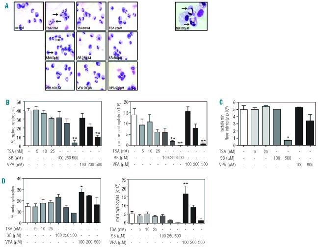
Figure 5.
Differential effects of HDAC inhibitors on terminal neutrophil differentiation. CD34+ cells were cultured in the presence of G-CSF for 17 days to induce neutrophil differentiation. Cells were cultured in either the absence or presence of TSA (5–25 nM), SB (100–500 μM) or VPA (100–500 μM). Cytospins were made and stained with May-Grünwald Giemsa solution (A). Arrows indicate hypersegmentation (5 nM TSA), hypergranulation and a ring shaped nucleus (100 μM SB). The right panel represents dysplastic neutrophil precursors (500 μM SB). Data are expressed as the percentage and absolute number (×106) of mature neutrophils (banded or segmented nuclei) (n=4) (B), lactoferrin expression (C) (n=2) and percentage and absolute number (×106) of metamyelocytes (D) (n=4). Error bars represent SEM (between experiments) *P<0.05, **P<0.01.

