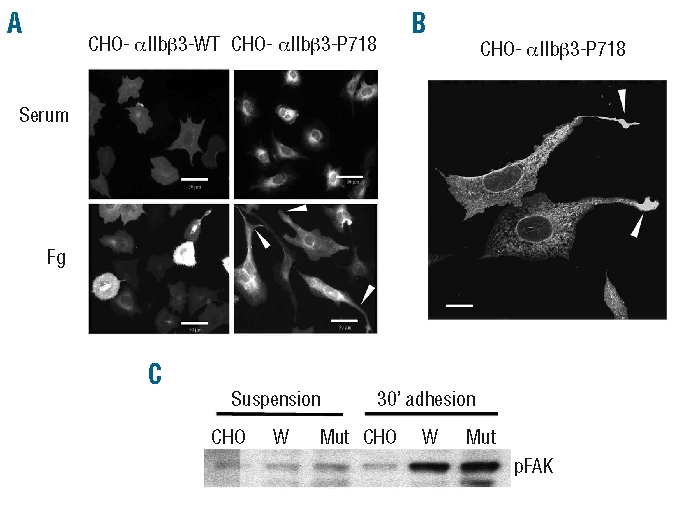
Figure 4.

Fibrinogen-mediated spreading of CHO cell transfectants. (A) Fluorescence microphotographs of CHO cell transfectants seeded for 16 h on fibrinogen or serum proteins. Cells were fixed, labeled with anti-β3 monoclonal antibody H1AG11 and examined in an epifluorescence microscope with a x40 objective. Arrowheads point to the extending processes in cells expressing mutant αIIbβ3-P718. Bars: 50 μm. (B) Arrows point to the swelling at the tip of the extending protrusion in CHO-αIIbβ3-P718 cells examined with a x63 objective. Bar: 25 μm. (C) Tyrosine phosphorylation of FAK in CHO cells. Non-transfected (CHO) or cells expressing wild-type (W) or mutant αIIbβ3-P718 were maintained in suspension or adhered to fibrinogen for 30 min. Cell lysates were analyzed by western blot. All the experiments were performed at least three times and similar results were obtained.
