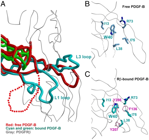
Fig. 4.
The PDGF-B conformational change upon PDGFRβ binding. (A) Comparison between the free PDGF-B (PDB code 1PDG) dimer (red) and the PDGFRβ-bound PDGF-B dimer (cyan for the protruding protomer and green for the receding protomer) shows that PDGFRβ-binding induces the structural organization of the previously disordered large L1 loop, and a rotation of the protruding L3 loop. (B) and (C) Comparison of PDGF-B Trp40 and its neighboring residues in the free form and receptor-bound form shows a 180° flipping of Trp40, as induced by its interactions with PDGFRβ Tyr205, Tyr207 and Phe136 (pink).

