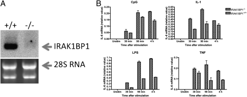
Fig. 1.
Increased IL-6 expression in IRAK1BP1 knockout mouse cells. (A) Northern blot analysis of IRAK1BP1 expression in knockout and wild-type fibroblasts. Ribosomal 28S RNA was used as loading control. (B) Embryonic fibroblasts from newly isolated IRAK1BP1-deficient or wild-type control embryos were activated with LPS (100 ng/mL), CpG (200 nm), IL-1 (20 ng/mL), and TNF (10 ng/mL) for different times followed by isolation of total RNA and quantitative PCR analysis of IL-6 mRNA accumulation. Data are representative of one (A) or three (B) independent experiments.

