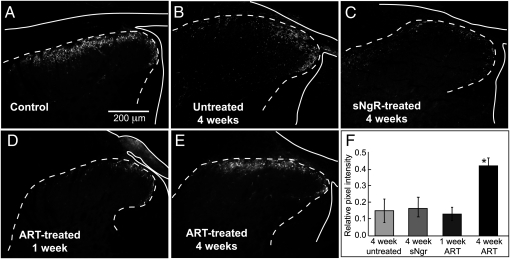
Fig. 3.
Expression of CGRP recovers after DR crush treated with ART, but not with sNgR. The outline of the cord and DR is indicated by a solid white line; the dotted line indicates the border between gray and white matter. (A) The normal projection of CGRP+ afferents was limited to upper laminae of the dorsal horn. (B and C) Four weeks after crush of five cervical DRs, CGRP expression fell to ~15% of normal in untreated cords (B) or with sNgR treatment (C). (D and E) Little recovery of CGRP expression was seen after 1 wk with ART treatment (D), but after 4 wk, expression recovered to ~40% of normal and was restricted to superficial laminae (E). (F) Quantitative assessment of recovery of CGRP expression in superficial laminae of untreated and sNgR or ART-treated cords with DR crush compared with uncrushed controls. The asterisk denotes significant difference from all other values (P < 0.001). Images in control panel (uncrushed axons) has been reversed horizontally in A, to facilitate comparison with projections of regenerated axons.

