Fig. 2.
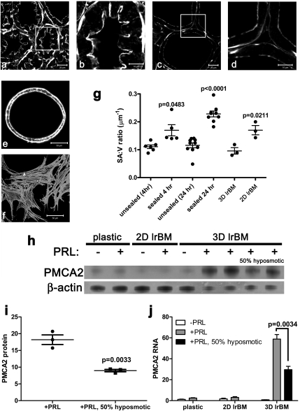
PMCA2 levels are associated with cell shape. Unsealed (A and B) and sealed (C and D) mammary glands, as well as 3D and 2D cultures of MMEC (E and F, respectively) were stained with Alexa-488-phalloidin to visualize microfilaments. Morphometric measurements (G) revealed that the surface area to volume ratio (SA:V) was significantly higher in sealed mammary glands and that the epithelial layer became thinner as milk accumulated over 24 h. Each point in G represents the measurement of the alveoli in one individual field-of-view. One-way ANOVA gave an overall P < 0.0001, and the Newman-Keuls multiple comparison posttest showed significant differences between unsealed and sealed glands (at 4 and 24 h) and between sealed glands at 4 vs. 24 h (unsealed 4 h n = 6; sealed 4 h, n = 5; unsealed 24 h, n = 10; sealed 24 h, n = 9). In addition, MMEC grown in 2D on lrBM were thinner than those grown in 3D (P = 0.0211, n = 3). The Student's t test P values are shown above the bars in graph g. Western blotting of 100 μg (lanes 1–5) or 50 μg (lanes 6–9) of total protein (H) and qRT-PCR (J) show that maximal PMCA2 expression in MMEC requires prolactin (PRL) and culture in 3D on lrBM. PMCA2 levels were reduced (P = 0.0033 for protein, P = 0.0034 for RNA) by mechanical deformation of 3D MMEC cultures with 50% hyposmotic treatment for 4 h (H–J). For protein, +PRL, n = 3; +PRL with 50% hyposmotic media, n = 3. For RNA, plastic –PRL, n = 7; plastic +PRL, n = 8; 2D lrBM −PRL, n = 8; 2D lrBM +PRL, n = 8; 3D lrBM −PRL, n = 3; 3D lrBM +PRL, n = 6; 3D lrBM +PRL with 50% hyposmotic media, n = 3. The mean ± SEM values are shown, with individual data points for G and I.

