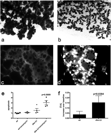
Fig. 3.
Lack of PMCA2 causes apoptosis of MECs during pregnancy. Mammary glands were taken from homozygous dfw-2J mice, which lack PMCA2 expression, and WT mice on day 18 of pregnancy. Alveoli were smaller in mammary whole mounts prepared from dfw-2J mice (B) compared with WT mice (A). TUNEL staining of WT (C) and dfw-2J (D) mammary glands at P18 revealed many more apoptotic nuclei in dfw-2J glands than in WT glands. Primary MMEC were cultured on lrBM with lactogenic media and treated with 5 μM ionomycin (Invitrogen) or vehicle for 16 h before analysis of apoptosis using the Cell Death ELISAPLUS(Roche) (E). WT MMEC in 3D lrBM culture were relatively resistant to 5 μM ionomycin-induced intracellular calcium stress (E), although in dfw-2J MMEC, ionomycin induced apoptosis (P = 0.0009; n = 4). The increased apoptosis in dfw-2J MMEC treated with 5 μM ionomycin is associated with a higher intracellular calcium level when compared with WT MMEC (F) (P = 0.0384, WT n = 122 cells in two experiments, dfw-2J n = 51 cells in two experiments). Cells were imaged while perfused with standard media for approximately 2 min to determine the baseline calcium levels, followed by perfusion with media containing 5 μM ionomycin. Data are expressed as mean percent increase in fluorescence over baseline. Error bars represent the mean ± SEM.

