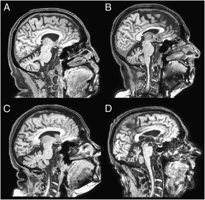
Fig. 3.
Structural MRI scans of SCA-6–induced cerebellar degeneration in four patients. (A) P27: male, 60 y, 3 y postonset, x 4. (B) P32: female, 62 y, 6 y postonset, x +4. (C) P07: female, 69 y, 10 y postonset, x −7. (D) P13: male, 74 y, 20 y postonset, x −3. x coordinates according to MNI convention.

