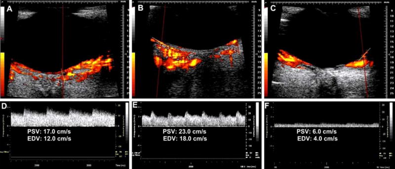
Fig. 3.

Combined power Doppler and spectral Doppler imaging of a long posterior ciliary artery (a, d), CRA (b, e), and the branch retinal artery (c, f). Blood velocity in the artery is represented by peak systolic velocity (PSV) and end diastolic velocity (EDV)
