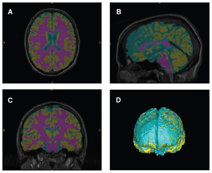
Fig. 1.
False colour of grey and white matter segmentation. Representative participant magnetic resonance imaging data following image registration and segmentation showing the axial (A), sagittal (B) and coronal (C) views, with 3-dimensional surface rendering (D). An atlas-based 3-channel tissue segmentation program (Expectation Maximization Segmentation) was used to segregate white matter (puce), grey matter (gold) and cerebrospinal fluid (blue).

