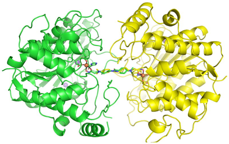
Figure 6.

The crystallographic dimer of the D101L Fe2+-HDAC8-M344 complex. Monomer A and monomer B are shown in green and yellow, respectively. Fe2+-coordinating residues and L101 are shown as sticks. Protein atoms are color coded as follows: carbon (green or yellow), oxygen (red), nitrogen (blue). Ions are colored as follows: Fe2+ (orange), K+ (purple). Inhibitor M344 atoms are colored as follows: carbon (green or yellow), nitrogen (blue), oxygen (red).
