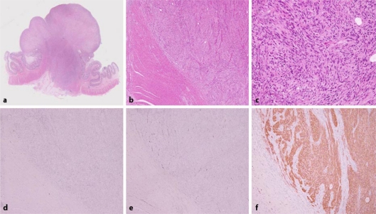
Fig. 2.
Histopathological examination findings. The majority of the upper portion of the lesion was replaced by granulation tissue (a). Spindle cells proliferated in a fascicular pattern (b). Nuclear palisading was observed in part (c). KIT (d), CD34 (e), desmin and smooth muscle actin were negative and S-100 (f) protein and NSE were positive in immunohistochemical staining.

