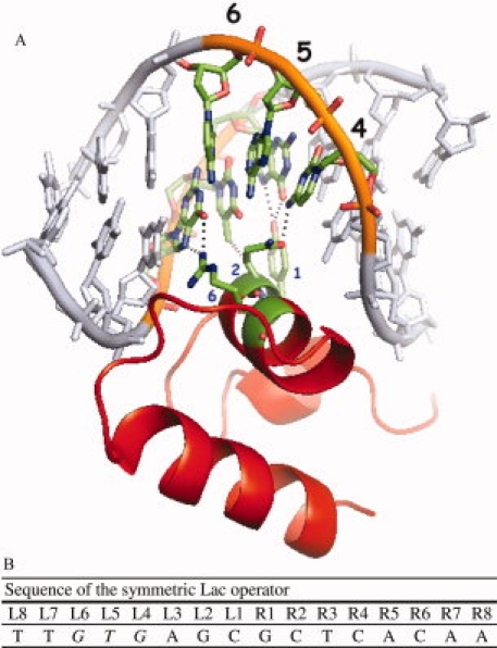
Figure 1.

(A) The structure of the Lac repressor bound to the symmetric operator. The recognition helix fits into the major groove of the DNA and residues 1, 2 and 6 of the recognition helix (Y17, Q18, R22) make specific interactions with operator positions 4, 5 and 6 of the operator (Bell and Lewis, 2000). Reprinted with permission from Oxford University Press, Daber and Lewis, Towards Evolving a Better Repressor, Protein Engineering Design and Selection, 2009, vol. 22, no. 11, p 673–683. (B) The symmetric Lac operator as identified by Sadler et al. (1983) and typically referred to as ‘symL (−1)’. “L” and “R” denote the left and right half-sites of the operator. An is available in the electronic version of the article. PRO389 Figure 1
