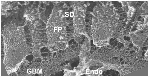
Figure 1. View of the glomerular capillary wall by freeze fracture deep-etch scanning electron microscopy.
The GCW consists of the diaphragm-less fenestrated endothelium (Endo), the GBM with its thick central layer (corresponding to the lamina densa by transmission EM), and podocyte FPs with bridging SDs. Note the thin strands connecting podocytes and endothelial cells to the GBM. Image provided by Dr. John Heuser, Washington University School of Medicine.

