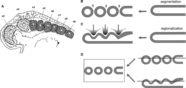
Fig. 2.
Is the head mesoderm of the lamprey segmented? (A) A parasagittal section of a young lamprey embryo published by Koltzoff (1901) (modified from de Beer 1937). The head mesoderm is colored light gray and real somites in the trunk are colored dark gray. In this section, numbering of the mesodermal “segments” starts from the yet undeveloped premandibular mesoderm (labeled “s1”) followed by the mandibular mesoderm (labeled “s2”). Thus, the first (real postotic) somite in the generic terminology is labeled “s4,” and “s3” in this figure corresponds to the sum of hyoid mesoderm and somite 0 (see Kuratani et al. 1999 for a definition of somite 0). At this stage, the head mesoderm appears to be “segmented” only between s2 and s3. (B) Schematic illustration of segmentation during somitogenesis in vertebrate embryos. (C) The regionalization process typical of the vertebrate head mesoderm. The unsegmented mesoderm is simply regionalized by surrounding structures and no real segmentation is evident. (D) A scheme showing that parasagittal sections cannot discriminate between segmentation and regionalization.

