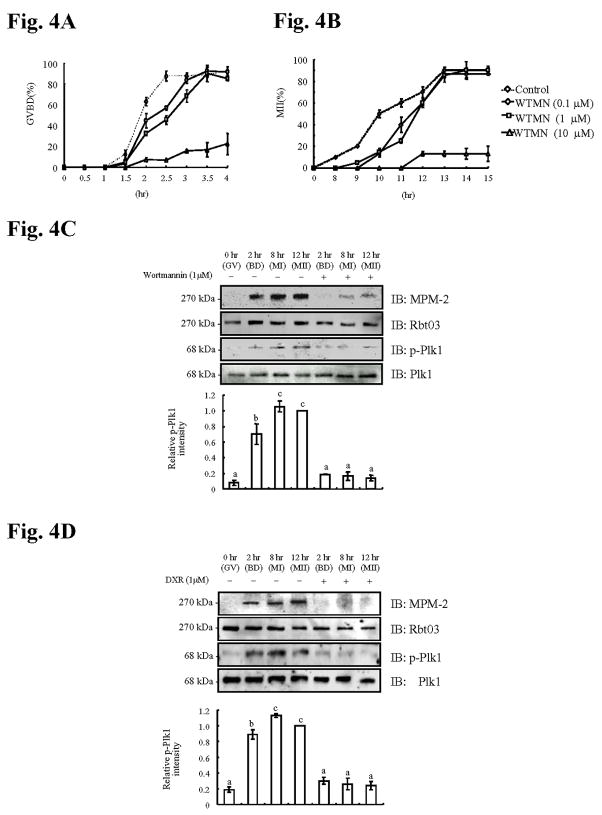
Fig. 4. Inhibition of Plk1 activity by Wortmannin (WTMN), or Doxorubicin (DXR) abolishes IP3R1 MPM-2 reactivity and delays progression to GVBD and MII.
Percentage of oocytes (control, 0.1, 1 or 10μM WTMN) that underwent progression to GVBD (A) and MII (B) during maturation; at least 30 oocytes were examined per concentration and time point. Immunoblotting of oocyte lysates collected at 0, 2, 8 and 12h ± 1μM WTMN (C) or ± 1μM DXR (D) were probed with MPM-2 (upper panel) antibody and p-Plk1 antibody (3th panel) and, after stripping, re-probed with IP3R1 antibody (Rbt03; 2nd panel) and Plk1 (lower panel), respectively. Relative p-Plk1 intensity is shown for both (4C and 4D) in the graphs below the immunoblotting panels.

