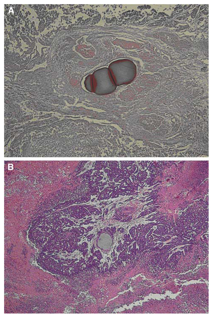
Fig. 6.
Pathologic images obtained at 7 days after treatment according to the treatment modalities. A A case treated with doxorubicin-loaded QuadraSphere microspheres shows near complete tumor cell death, whereas B a case treated with bland embolization shows residual viable cells around the embolized area. Note that the pink color of the QuadraSphere microspheres containing doxorubicin at 7 days after treatment indicates that doxorubicin is still present in the micropshere (A), in contrast to the gray color of plain QuadraSphere microspheres (B) (Color figure online)

