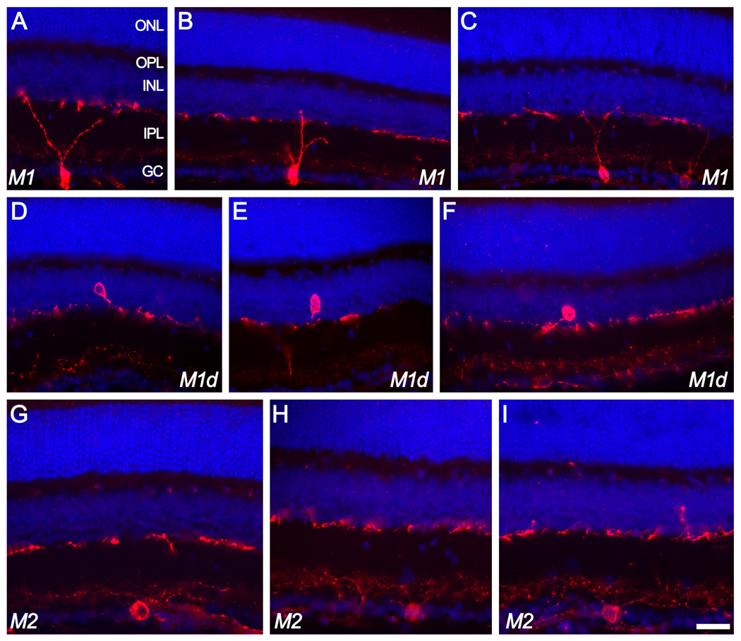
Figure 1.
Ganglion cells of the M1 and M2 types as revealed in vertical sections of the mouse retina immunostained for melanopsin (red). A nuclear counterstain (DAPI; blue) reveals cellular laminae. Top two rows of images (A–F) illustrate melanopsin-immunoreactive cells of the M1 type, characterized by dendritic arborizations in the outer melanopsin immunoreactive plexus lying at the boundary between inner plexiform and inner nuclear layers. Some of these M1 cells were conventionally placed with somata in the ganglion cell layer (top row; A–C) while others (“M1d”) had somata displaced to the inner nuclear layer (middle row; D–F). The fainter inner (M2) plexus is barely detectable in some of these images. M2 cells (bottom row; G–I) had conventionally placed somata and laterally spreading dendrites entering the inner (M2) plexus of immunopositive dendrites. Scale bar: 30 µm. ONL=outer nuclear layer; OPL=outer plexiform layer; INL=inner nuclear layer; IPL=inner plexiform layer; GC=ganglion cell layer.

