Figure 1.
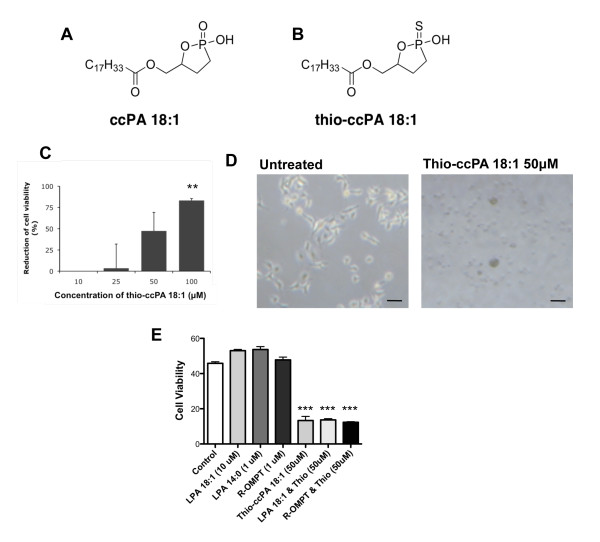
Thio-ccPA 18:1 reduces the viability of B16F10 cells in vitro. (A) Chemical structure of ccPA 18:1 and (B) thio-ccPA 18:1. (C) B16F10 cells were treated with increasing concentrations (10-100 μM) of thio-ccPA 18:1 and analyzed for cell viability after 48 h. The graph presents the data as the percentage of reduction in cell viability (% of PBS control). **p < 0.01 vs. control by Bonferroni's t-test and analysis of variance. (D) B16F10 cells were either untreated (control) or treated with thio-ccPA 18:1 (50 μM). Images demonstrate the difference in B16F10 cell morphology after 48 h treatments with 50 μM thio-ccPA 18:1. (E) B16F10 cells were either untreated (control) or treated with LPA 18:1 (10 μM), LPA 14:0 (1 μM), R-OMPT (1 μM), thio-ccPA 18:1 (50 μM) or a combination of these as shown. Cell viability was assessed after 48 h. ***p < 0.001 vs. control by Bonferroni's t-test and analysis of variance.

