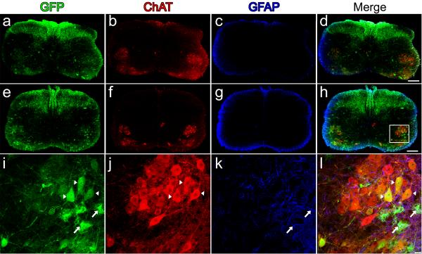
Figure 1.
Intravenous injection of AAV9 leads to widespread neonatal spinal cord transduction. Cervical (a–d) and lumbar (e–l) spinal cord sections ten-days following facial-vein injection of 4×10¹¹ particles of scAAV9-CB-GFP into postnatal day-1 mice (n=8). GFP-expression (a,e,i) was predominantly restricted to lower motor neurons (a,e,i) and fibers that originated from dorsal root ganglia (a,e). GFP-positive astrocytes (i,l, arrows) were also observed scattered throughout the tissue sections. Lower motor neuron and astrocyte expression were confirmed by co-localization using choline acetyl transferase (ChAT) (b, f,j) and glial fibrillary acidic protein (GFAP) (c,g,k), respectively. Merged images of GFP, ChAT and GFAP (d,h,l). A z-stack image (i–l) of the area within the box in h, shows the extent of motor neuron (arrow heads) and astrocyte (arrows) transduction within the lumbar spinal cord. Scale bars, 200 μm (d,h), 20 μm (l).

