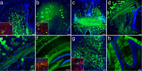
Figure 3.
Widespread GFP-expression 21-days following intravenous injection of 4×10¹¹ particles of scAAV9-CB-GFP to postnatal day-1 mice. GFP localized in neurons and astrocytes throughout multiple structures of the brain (n=6) as depicted in: (a) striatum (b) cingulate gyrus (c) fornix and anterior commissure (d) internal capsule (e) corpus callosum (f) hippocampus and dentate gyrus (g) midbrain and (h) cerebellum. All large panels show GFP (green) and dapi (blue) merged images. Insets of selected regions show high magnification merged images of GFP (green), NeuN (red) and GFAP (blue) labeling. Schematic representations depicting the approximate locations of each image throughout the brain are shown in (Supplementary Fig. 6). Higher magnification images of select structures are available in (Fig. 4). Scale bars, 200 μm (a); 50 μm (e); 100 μm (b–d,f–h)

