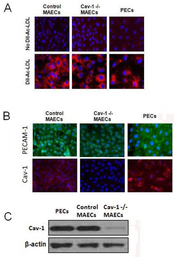
Figure 1.
Characterization of endothelial cells. (A) Fluorescent microscopy of mouse and porcine endothelial cells demonstrating uptake of Dil-Ac-LDL labeling. Pictures were taken at 400× magnification. (B) Fluorescent microscopy of mouse aortic endothelial cells (MAECs) and porcine endothelial cells (PECs) that express PECAM-1 (FITC, green) and caveolin-1 (Cav-1) (Texas Red, red). (C) Caveolin-1 protein expression in mouse and porcine endothelial cells determined by SDS-PAGE and Western blot analysis. β-actin was used as loading control.

