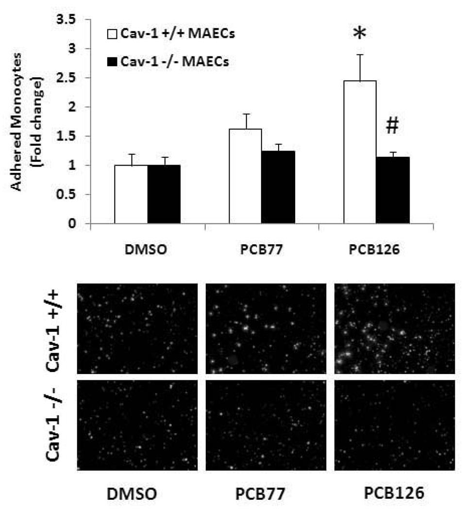
Figure 6.
Monocyte adhesion to MAECs exposed to coplanar PCBs. MAECs were exposed to PCB77 or PCB126 at 2.5 µM for 16 h. Human THP-1 monocytes were activated with TNF-α and loaded with the fluorescent probe calcein. Activated and calcein loaded monocytes were added to endothelial monolayers for adhesion. After washing, adhered monocytes were counted using a fluorescent microscope. Results represent the mean ± SEM, with n=3. Experiments were repeated a minimum of three times. Representative microphotographs show adhered monocytes on endothelial cell monolayers. *Significantly different compared to DMSO control. #Significantly different compared to corresponding control C57BL/6 mouse endothelial cells.

