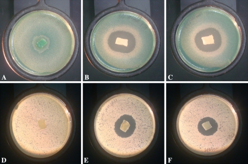
Fig. 3A–F.
(A–C) Agar plates with Pseudomonas aeruginosa. (A) Saline-loaded chitosan sponge control and (B–C) chitosan sponges loaded with amikacin. No inhibition of growth was seen with the saline-loaded control. (D–F) Agar plates with Staphylococcus aureus. (D) Saline-loaded chitosan sponge control and (E–F) are chitosan sponges loaded with vancomycin. No inhibition of growth was seen with the saline-loaded control. The images were taken at the 48-hour time point.

