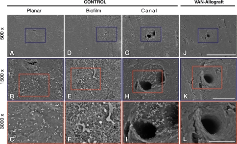
Fig. 7A–L.
SEM assessment of colonization is shown. Control surfaces were abundantly colonized with bacteria (CONTROL) after 12 hours. At low magnification on the planar surfaces (A), small white dots are evident that, at higher magnifications (B–C), resolve into multiple colonies of bacteria that populate the surface in grape-like clusters. On other areas, reticulated biofilm (D) is evident which at higher magnification (E–F) can be distinguished as a thick layer of slimy material with multiple small spherical bacteria (diameter 0.3–1 μm) representing biofilm entrenched S. aureus colonies. Natural topographic niches such as Haversian and Volkmann’s canals (G) at low magnifications showed no apparent colonization. However, at higher magnifications, they were consistently filled with S. aureus (H) that at 3000x (I) were seen deep into these canals. The surface of the VAN-allograft appeared free of bacterial colonies on all surfaces at any magnification (J–L). Scale bars: A, D, G, J = 200 μm; B, E, H, K = 50 μm; C, F, I, L = 30 μm.

