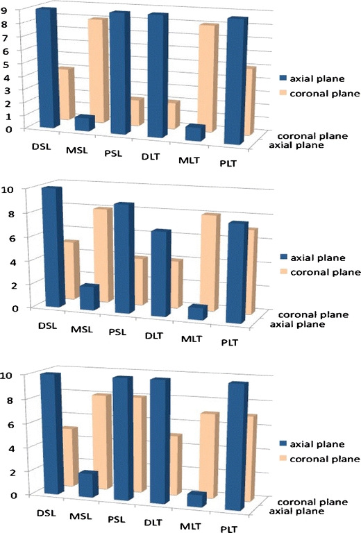
Fig. 7.

Detectability of different regions of the scapholunate and lunotriquetral ligaments by MR arthrography in radial deviation (top), ulnar deviation (middle) and neutral position (bottom). Y-axis number of ligaments observed, X-axis portions of the ligaments. DSL dorsal scapholunate, MSL membranous scapholunate, PSL palmar scapholunate, DLT dorsal lunotriquetral, MLT membranous lunotriquetral, PLT palmar lunotriquetral
