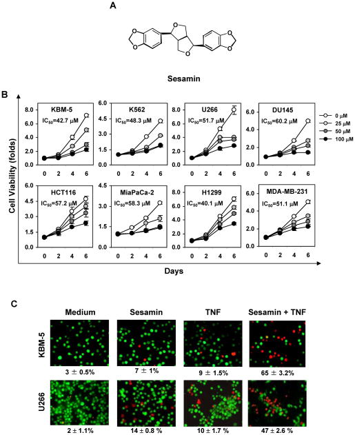
FIGURE 1.
(A) Chemical structure of sesamin. (B) Sesamin suppresses tumor cell proliferation. Cells were incubated with 0, 25, 50, and 100 μM sesamin for different days. Cell proliferation was then analyzed by the MTT method as described under “Materials and Methods.” (C) Sesamin potentiates TNF-induced cytotoxicity. KBM-5 (1 × 10⁶) or U266 (1 × 10⁶) cells were incubated with 100 μM sesamin for 12 h and then incubated with 1 nM TNF for 24 h. The cells were stained with a Live/Dead assay reagent for 30 min and then analyzed under a fluorescence microscope as described under “Materials and Methods.”

