Figure 1.
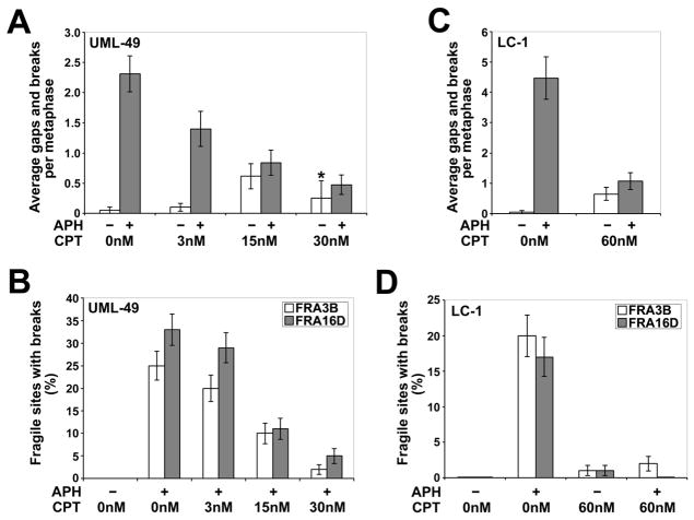
Low doses of CPT reduce the frequency of APH-induced common fragile site breaks. (A) Average total chromosomal gaps and breaks per cell in UML-49 cells after 18 h in the presence (gray) or absence (white) of 0.4 μM APH and 0 to 30 nM CPT; n= 80 metaphases for each data set except for cells treated with only 30 nM CPT (asterisk), in which case n=8 due to very few metaphases being present for scoring. Error bars indicate the 95% confidence interval. (B) Frequency (%) of gaps and breaks at specific fragile sites FRA3B and FRA16D in UML-49 cells after 18 h in the presence or absence of 0.4 μM APH and 0 to 30 nM CPT; n= 96 sites examined. Fragile sites were identified by G-banding. Frequency of fragile-site induction is presented as the percentage of chromosome 3 or 16 homologs with breaks at FRA3B (white) or FRA16D (gray), respectively. Error bars indicate the 95% confidence interval. (C) Average total chromosomal gaps and breaks per cell in LC-1 cells after 18 h in the presence (gray) or absence (white) of 0.3 μM APH and 0 or 60 nM CPT; n= 80 metaphases for each data set. Error bars indicate the 95% confidence interval. (D) Frequency (%) of gaps and breaks at specific fragile sites FRA3B and FRA16D in LC-1 cells after 18 h in the presence or absence of 0.3 μM APH and 0 or 60 nM CPT; n= 100 sites examined. Fragile sites were identified by G-banding. Frequency of fragile-site induction is presented as the percentage of chromosome 3 or 16 homologs with breaks at FRA3B (white) or FRA16D (gray), respectively. Error bars indicate the 95% confidence interval.

