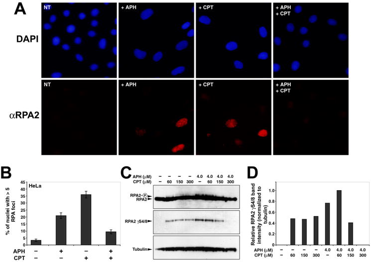
Figure 4.
CPT reduces APH-dependent RPA2 nuclear focus formation and RPA2 phosphorylation. (A) Examples of immunocytochemistry performed with a RPA2-specific antibody in HeLa cells following 24 hour treatment with 4.0μM APH, 300nM CPT, or both. Nuclei are stained with DAPI. (B) Quantitation of the number of nuclei with >5 nuclear RPA2 foci after drug treatments (n > 200 nuclei per treatment). (C) Western blots showing the induction by APH of RPA2 phosphorylation as evidenced by a gel shift (panel 1) and by detection of phosphorylation on serines 4 and 8 (panel 2). Addition of CPT reduces the APH-depended phosphorylation of RPA2 in a dose-dependent manner. Tubulin is included as a loading control. Concentrations of APH and CPT, added for 24 hours, are indicated. A 10 μM CPT treatment for 2 hours was included as a positive control. (D) Quantitation of RPA2 phosphorylation in (C), normalized to tubulin.

