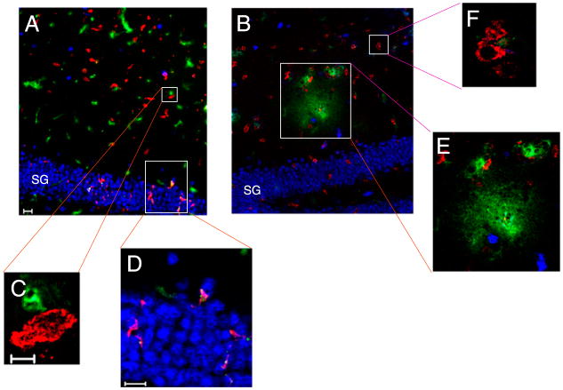
FIGURE 2.
Brain-infiltrating CD8+ cells are in close proximity to neurons and blood vessels during early CNS vascular permeability. Mouse brains isolated from 7-d TMEV-infected mice were immunostained for NeuN (blue) and CD8a protein (red). FITC-albumin (green) was administered i.v. 1 h prior to brain harvest and denotes vessels as well as vascular leakage. A, At 0 h, CD8+ cells are detected in the hippocampus (original magnification ×20) in close proximity to (C) vessels (original magnification ×100), and (D) neurons of the SG of the hippocampal dentate gyrus (original magnification ×40). B, FITC-albumin leakage is dorsal to the SG 4 h postadministration of VP2121–130 (original magnification ×20). In this region, CD8+ cells are found near (F) intact vessels (original magnification ×100) and (E) areas with FITC-albumin leakage (original magnification ×40). Shown is a section from a representative mouse of four mice analyzed in each group. Scale bars are as follows: in A, 20 μm for A and B; in C, 5 μm for C and F; in D, 20 μm for D and E.

