FIGURE 4.
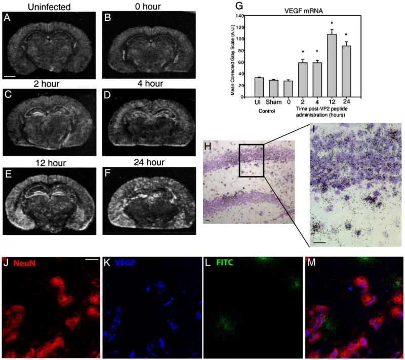
Neurons are a major source of VEGF expression in the CNS after induction of CD8 T cell-mediated CNS vascular permeability. Shown are representative coronal sections of mice at 0, 2, 4, 12, and 24 h postadministration of VP2121–130 peptide (n = 4 animals per time point). All sections were analyzed for VEGF mRNA and protein expression. In situ hybridization reveals areas of the brain that express VEGF mRNA in (A) uninfected, (B) 0 h, (C) 2 h, (D) 4 h, (E) 12 h, and (F) 24 h post-VP2121–130 peptide administration. G, VEGF mRNA labeling was quantified in the ipsilateral SG of the hippocampal dentate gyrus. H and I, Emulsion autoradiographs with cresyl violet counterstaining of the hippocampal dentate gyrus demonstrate substantial VEGF mRNA patchy neuronal labeling in the SG and subjacent hilar region (H, original magnification ×20; I, original magnification ×40). Also shown is confocal microscopy of (J) NeuN, (K) VEGF, (L) FITC-albumin in hippocampus 12 h postadministration of VP2121–130 peptide (original magnification ×100). NeuN immunostaining colocalizes VEGF cytokine as shown merged in (M). Scale bars are as follows: (A) 1300 μm for A–F, (H) 20 μm, (I) 20 μm, and (J) 10 μm for J–M. *Denotes statistical significance with p < 0.05 when compared with 0 h.

